


Precision oncology – treatment of canine osteosarcoma
Precision oncology – chemotherapeutic drug delivery with nanoparticles as latest improvement in the treatment of canine osteosarcoma
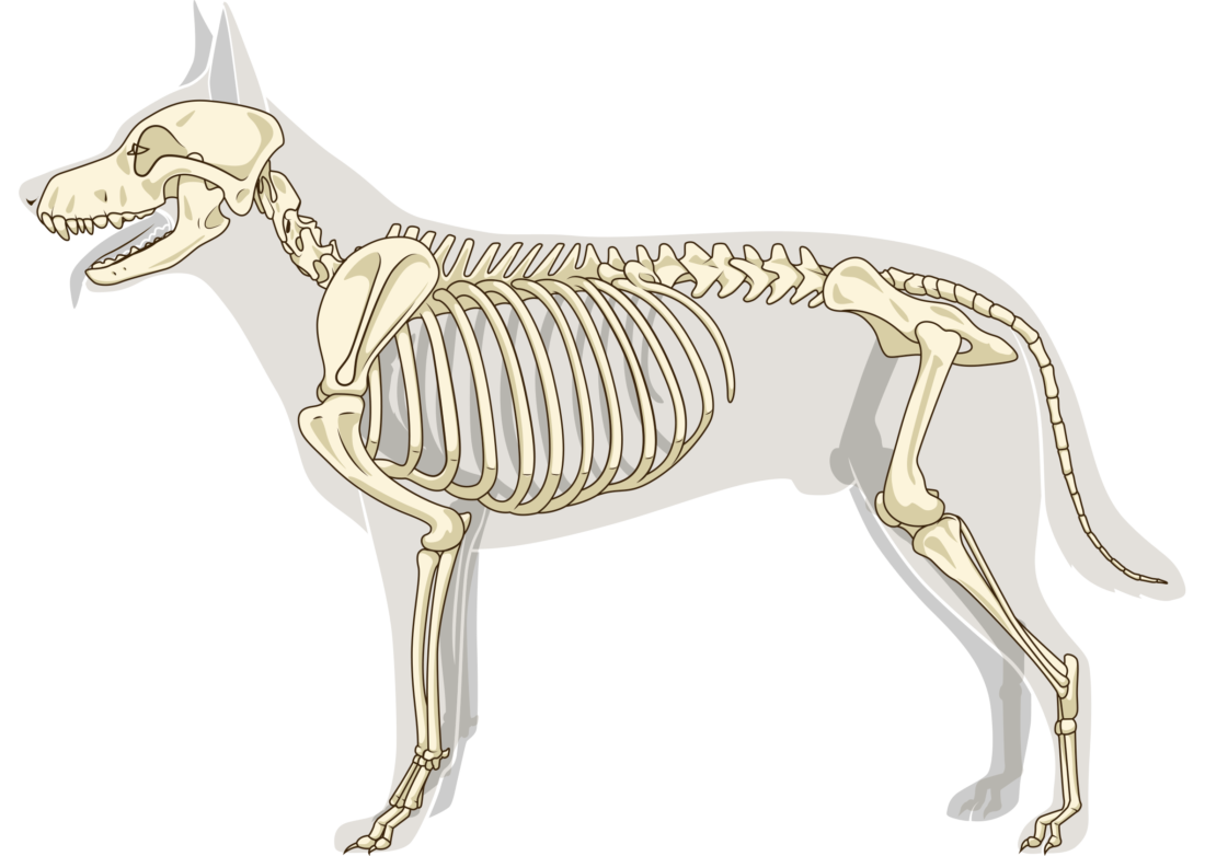
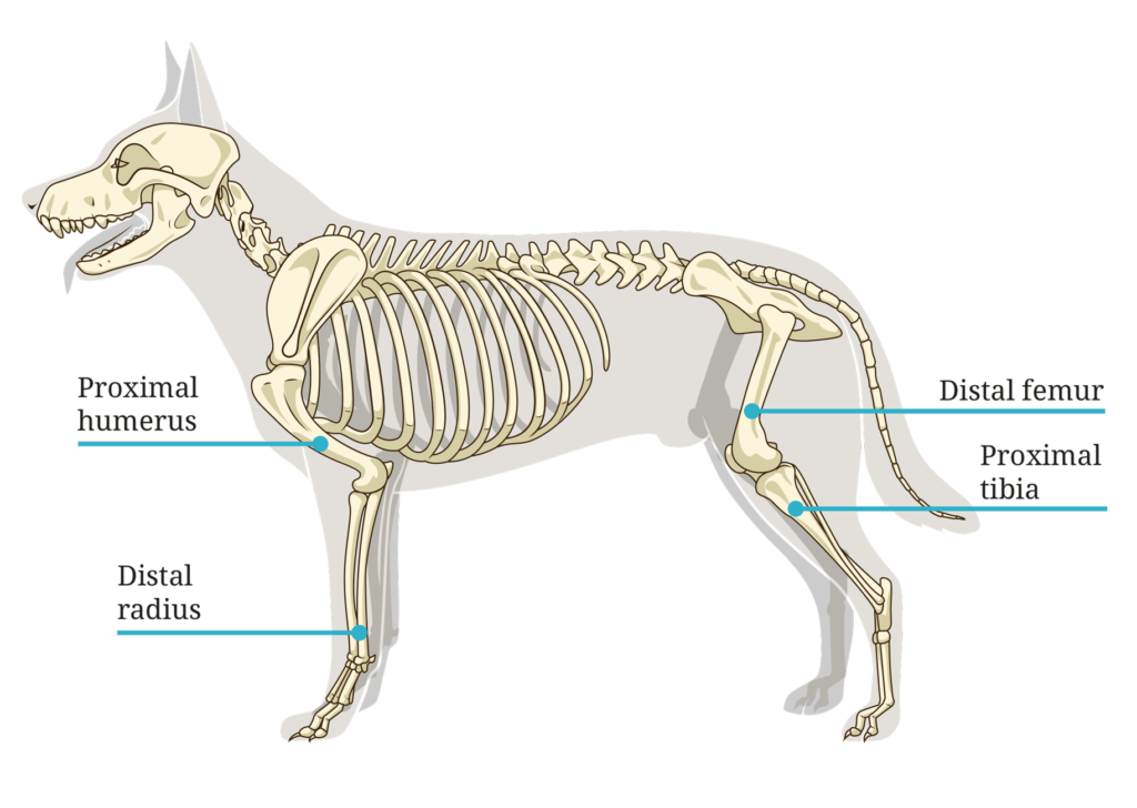
Osteosarcoma is an agressive bone cancer caused by uncontrolled differentiation of mutated osteoblastic cells that leads to malignant osteoid. Osteosarcoma is the most frequent form of primary bone cancer in dogs. It commonly occurs in larger middle aged (6 to 10 years old) breeds such as German Shepherds, Boxer, Rottweiler or Doberman Pinscher. Different studies revealed several risk factors for developing osteosarcoma such as a higher frequency in males than females or castration. More than half of the canine osteosarcoma cases develop the malignancy in the appendicular skeleton and with less frequency in the axial skeleton or the extraskeletal muscles. This form of bone cancer has a very high ability to metastasize and about 80% of affected dogs die due to lung metastasis. The location of the primary tumor increases the risk of developing metastasis. Specifically, tumors localized at the proximal humerus and distal femur or the proximal tibia have a higher metastasis ability leading to a higher mortality (1).
Due to the high probability of metastasis and other complicating factors, treatment of canine osteosarcoma remains a challenge for veterinary physicians. But which are the treatment options up to now? Once osteosarcoma is diagnosed, there are different treatment options including surgery, limb amputation or limb-sparing surgery, as well as radio – and chemotherapy (1). A limb amputation is the most performed treatment method which gives pain relief and prolongs survival. A long term retrospective study in dogs with stage III osteosarcoma showed that a palliative treatment with a combination of surgery, radiation and chemotherapy led to a statistically significant longer survival time (130 days) compared to treatment with surgery alone (3 days) or when treated with surgery and chemotherapy (78 days). The same study also revealed that dogs with osteosarcoma and bone metastasis showed a better survival time than affected dogs with soft tissue metastasis (2).
The limb-sparing surgery represents an alternative treatment option to amputation of the cancerous bone. The method has been applied in dogs with osteosarcoma for several years. Hereby, either a metal implant or a cortical allograft is used to replace the affected bone segment. In order to perform a limb-sparing surgery a large bank of sterilely harvested cadaveric cortical bone and experienced surgeons are required. Unfortunately, after the surgery, nearly 50% of the treated dogs suffer from infections (30 – 50% of the cases) or the implant is rejected (20 – 40% of the cases). Additional disadvantages of this method are chronic pain or poor limb use finally leading to amputation. But how could infections or implant rejection be circumvented and the affected bone be preserved? N. Ehrhart studied an alternative treatment option, the longitudinal bone transport osteogenesis (BTO). BTO is a technique to reconstruct large segmental bone defects caused by trauma or infection. A piece of bone is separated from the affected bone by osteotomy. This bone segment serves as the transport element. With the help of an external ring construct, the bone segment is moved slowly along the bone defect while new bone is regenerated. The newly created bone has an increased resistance to infection because of a constant blood supply. N. Ehrhart could show in her study that after treatment limb function could be recovered good to excellent in 7 out of 9 dogs. Eventhough allograft limb salvage seems to be the simpler technique, BTO represents several advantages such as that the defect is replaced by living, autogenous bone and the reduction of infections and allograft resorption. Besides, BTO showed some promising long-term outcomes (3).
The BTO technique has been further improved by C. T. Jehn and colleagues who applied the transverse ulnar bone transport osteogenesis. Here, a fixator construct with reeling or linear motors transports an ulnar segment transversely into the defect created by excising the distal 50% of the ipsilateral radius. A longitudinal osteotomy of the adjacent ulna was created and the segment transported across the radial defect. The study in one live dog showed that the two created constructs transported the bone segment effectively and the dog produced viable regenerate bone. The researchers could conclude from this study that transverse ulnar bone osteogenesis is a potential method for limb salvage. The advantage compared to BTO is the substantially less process time because of the shortening of the transport distance (4).
Treatment of osteosarcoma has been observed to be more effective when surgery is combined with chemotherapy. Advances in chemotherapeutic drug treatment have also been made during the last years. A researcher team around Jianjun Chen and Timothy Fan recently published their results testing a new targeted chemotherapeutic drug delivery method for the treatment of osteosarcoma in dogs. They produced and tested specially engineered bone-seeking nanoparticles carrying doxorubicin an coated with pamidronate. Pamidronate is a drug that is attracted by areas of active bone repair, ideal for the treatment of osteosarcoma where bone tissue is destroyed and reconstructed continuously. Chen and his researchers first performed studies in vitro and in mice in order to elucidate the functionality and efficacy of their delivery system. They could show that the nanoparticle constructs were attracted in vitro by dishes mimicing the surface of bones. In mice they could demonstrate that this treatment slowed down tumor growth and reduced bone loss. Later they performed a phase I clinical study with nine dogs with osteosarcoma. The dogs received the nanoparticle construct intravenously. It was demonstrated that the nanoparticles accumulated in the bone tumor without any sgns of toxicity for the dog. After three weeks of treatment the dogs limbs were amputated and each tumor showed dying tumor tissue in the bone as a result of the drug treatment. This study could show that the new treatment method was effective (5).
In summary, continuous improvements are investigated for the treatment of dogs with osteosarcoma in order to prolong their lives and give pain relief. The demonstrated studies towards a better and more effective treatment for this type of cancer are not only advantageous for the veterinary medicine but are also an important model for the treatment of humans.
References:
1 M. Szewczyk, R. Lechowski, K. Zabielska. What do we know about canine osteosarcoma treatment? – review. Vet Res Commun, 2015, 39: 61-67.
2 S. E. Boston, N. P. Ehrhart, W. S. Dernell, M. Lafferty, S. J. Withrow. Evaluation of survival time in dogs with stage III osteossarcoma that undergo treatment: 90 cases (1985 – 2004). Javma, 2006, 228, 12.
3 N. Ehrhart. Longitudinal bone transport for treatment of primary bone tumors in dogs: Technique description and outcome in 9 dogs. Veterinary surgery, 34: 24-34, 2005.
4 C. T. Jehn, D. D. Lewis, J. P. Farese, E. A. Ferrell, W. G. Conley, N. Ehrhart. Transverse ulnar bone transport osteogenesis: A technique for limb salvage for the treatment of distal radial osteosarcoma in dogs. Veterinary surgery, 36: 324 – 334, 2007.
5 Q. Yin, L. Tang, K. Cai et al. Pamidronate functionalized nanoconjugates for targeted therapy of focal skeletal malignant osteolysis. Pnas.
![]()
No Comments